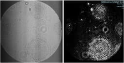
Digital inline holographic microscopy (DIHM): (from Bochdansky,et al (2017) JMS)
Details of the DIHM were published in Bochdansky et al. (2013). Briefly, a laser beam is focused on a 9 um single-mode optical fiber that serves as a small but intense point source of light. The expanding beam intercepts particles that create interfering shadow images on the adjacent screen of a high-resolution (4.2 megapixel) charge-coupled device (CCD) camera without a lens. The camera was connected to an eBOX530-820-FL1.6G-RC computer (Axiomtek) with a Gb LAN cable; images were recorded on a 750 GB hard disk at a frame rate of ~7-12 images per second. When the laser beam intercepts a structure, a portion of the image beam scatters and interferes with the light of the primary beam in a predictable pattern. This raw image represents a hologram that can then be reconstructed by applying the Kirchhoff–Helmholtz transform (Xu et al., 2001) in commercially available reconstruction software (Octopus, 4-Deep Inwater Imaging, formerly Resolution Optics). Being lens-less, the advantage of this method is that anything in the 7-cm long image beam can be reconstructed without having to adjust the focus on the object. The entirety of the image beam volume (i.e., 1.8 ml in this configuration) can be reconstructed in this fashion, and thus explores orders of magnitude more volume than any lens-based system would at the same resolution. Reconstruction of the images and analysis (particle quantities, sizes, and type) were performed manually as no reliable image reconstruction and analysis system currently exists for this custom-built DIHM. The DIHM is well suited to detect hard structures (e.g., silica, chitin, calcium carbonate, strontium sulfate) to a resolution as small as 5 um, and reliably images particles of any composition from 50 um to ~8 um in the image volume (Bochdansky et al., 2013). The DIHM does not "see" transparent exopolymers (TEP), which can only be inferred from the distribution of finer particles suspended in that matrix. Even at speeds of 1.5 m s-1 through the water, our instrument yields sharp images (Bochdansky et al., 2013, 2017).
NOTE: Phaeocystis colonies, because of their dense structure, did not reconstruct well (Fig. 3); however, they have a very characteristic shape and texture even in the unreconstructed holograms (Fig. 1) that we were able to verify in tests with laboratory cultures of P. antarctica. Consequently, we were able to perform a detailed analysis on Phaeocystis colonies on all casts through all depths (including the surface mixed layer).
Fig. 1. Four Phaeocystis antarctica colonies in a single unreconstructed hologram (a), and after reconstruction of one colony (b). The image volume of an individual hologram is 1.8 ml, but the reconstruction can only visualize a specific image plane within that volume. We concluded that Phaeocystis colonies were sufficiently distinguishable and unique that unreconstructed images could be used for quantification. Poor reconstruction of Phaeocystis colonies makes exact size determination unreliable but colony diameters in field collections in the Ross Sea range from approximately 10 to 400 um (Mathot et al., 2000). DOI: 10.4319/lom.2013.11.28


